
How Leqembi and Kisunla Fight Alzheimer's in Different Ways
When I was diagnosed early, I counted myself among the lucky ones. Early detection opened a small window of choice, and with it, the possibility of hope. Two new treatments, Leqembi and Kisunla, now offer a chance to slow Alzheimer’s progression for those in the early stages. Choosing between them felt like standing at a fork in the road; one that could shape how much of me I get to keep. Below is the information I wish I had known at the time, July 2024.
Understanding Alzheimer's Disease
When I was diagnosed with Alzheimer's disease, I thought the goal of treatment was simple: remove the amyloid plaque from my brain.
By 2026, I had learned something that completely changed my understanding of Alzheimer's.
Scientists now believe that the large amyloid plaques seen on PET scans are not the first or even the most damaging form of beta-amyloid. The disease begins years earlier with tiny, invisible clusters of beta-amyloid called oligomers. These microscopic protein clumps are now thought to be among the earliest and most toxic drivers of Alzheimer's disease.
To understand today's treatments, it helps to understand how beta-amyloid changes over time.
Every brain naturally produces a small protein fragment called beta-amyloid. Under healthy conditions, these fragments are continuously cleared away. As we age, and especially in people who develop Alzheimer's disease, some of these protein fragments are not removed efficiently. Over many years they gradually accumulate, particularly around the brain's waste-clearing system near the blood-brain barrier.
As these proteins build up, they begin folding into abnormal shapes. Instead of floating freely, they become sticky and attach to one another.
The process happens in stages:
· One beta-amyloid molecule is called a monomer.
· Two joined together form a dimer.
· Several molecules create an oligomer.
· Oligomers grow into protofibrils.
· Protofibrils become fibrils.
· Fibrils eventually collect into the amyloid plaques associated with Alzheimer's disease.
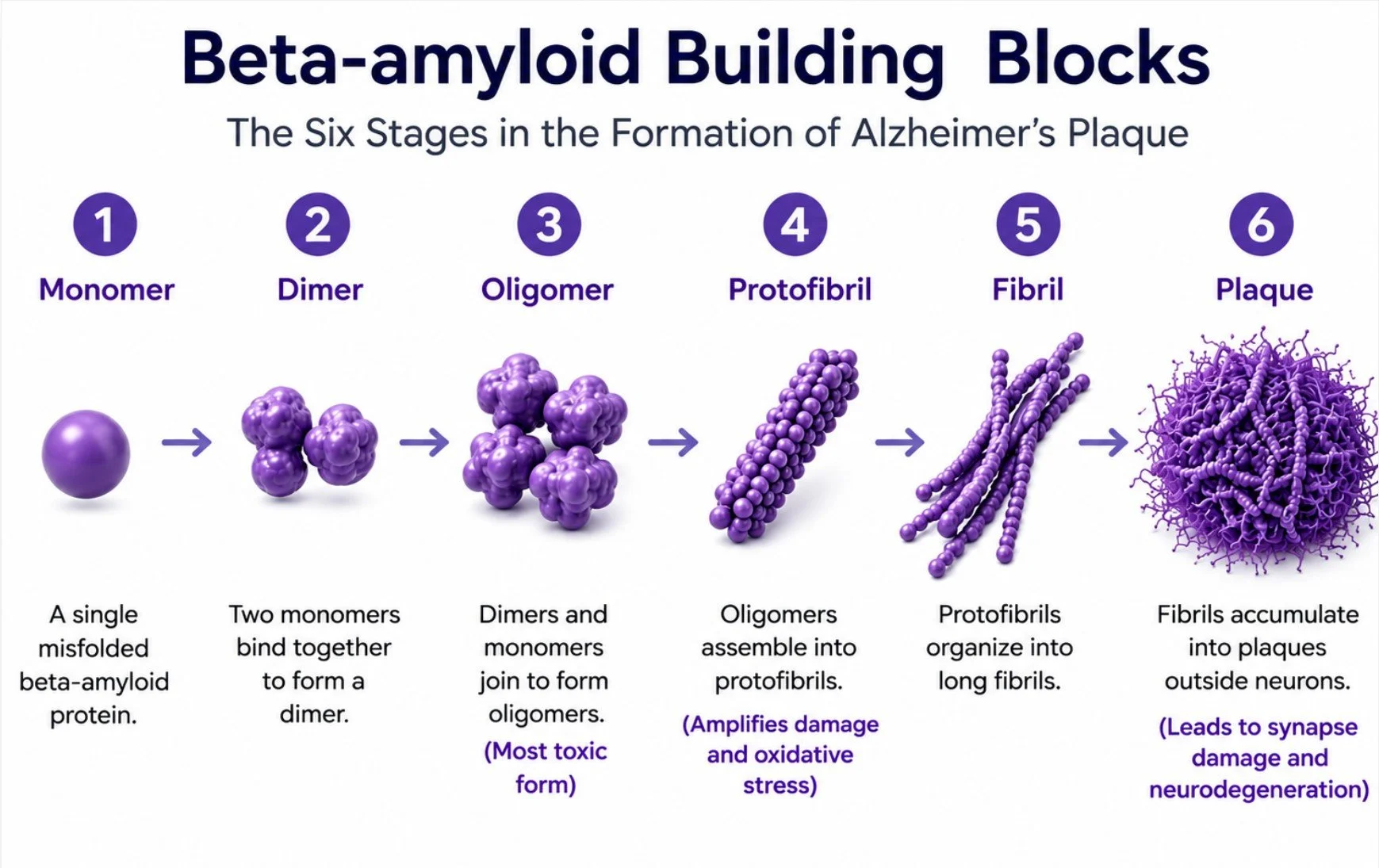
For many years, scientists believed plaques and tau tangles caused most of the damage. Today, research suggests that the much smaller oligomers may be the first "bad actors" in the disease.
Think of individual beta-amyloid molecules as tiny drops of honey floating through the brain. Alone, each drop is harmless. But when several drift together, they merge into a sticky blob called an oligomer. These tiny clumps are small enough to travel throughout the brain, attaching themselves to synapses the microscopic connections where neurons communicate and where memories and neural pathways are formed.
Unlike plaques, which remain relatively stationary once they form, oligomers continue moving from synapse to synapse, disrupting communication years before plaques become visible on a brain scan.
The Brain's Balancing System
While researching Alzheimer's, I discovered another fascinating part of the disease that helped explain why oligomers are so destructive.
Your brain depends on two chemical messengers that constantly work together.
Glutamate is the brain's accelerator. It excites nerve cells, allowing us to think, learn, remember, and react quickly.
GABA is the brain's brake. It calms brain activity, helps us concentrate, promotes restful sleep, and prevents neurons from becoming overstimulated.
Every thought, memory, movement, and conversation depends on these two chemicals remaining in balance.
Imagine driving a car. Glutamate presses the gas pedal. GABA applies the brakes. Neither one is "good" or "bad." You need both working together to reach your destination safely.
How Alzheimer’s Disrupts the Balance
Researchers have discovered that oligomers attack this delicate balance very early in the disease. They stimulate neurons to release too much glutamate, interfere with the brain's ability to remove the excess, and weaken GABA's calming influence. The result is a brain that is constantly pressing the accelerator while the brakes gradually begin to fail.
Over time, this excessive stimulation injures synapses, disrupts communication between neurons, and eventually causes neurons to malfunction and die. Scientists now believe these changes begin many years before noticeable memory loss and well before large amyloid plaques appear.
As oligomers continue to grow, they become protofibrils, then fibrils, and finally the amyloid plaques that accumulate throughout the brain. At the same time, the chronic stress created by toxic beta-amyloid, inflammation, and plaque formation causes another important protein tau to change.
Healthy tau normally acts like the railroad ties that stabilize the microscopic tracks inside neurons, allowing nutrients, energy, and chemical messages to travel efficiently throughout the cell. Under chronic stress, tau proteins detach from these tracks, fold into abnormal shapes, and begin sticking together.
Just as individual beta-amyloid molecules join together to form oligomers, protofibrils, fibrils, and eventually plaques, individual tau proteins also join together into small toxic tau oligomers, then fibrils, and finally the neurofibrillary tangles found inside dying neurons.
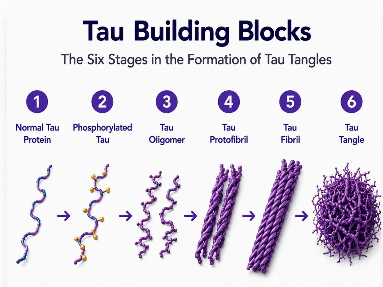
As tau tangles grow, they block the neuron's internal transportation system, starving the cell of nutrients and preventing it from communicating effectively. Eventually the neuron dies, and toxic tau fragments can spread to neighboring neurons, where the destructive process begins again.
None of this means that plaques are unimportant. Removing amyloid plaque remains one of the greatest advances in Alzheimer's treatment. Plaques serve as a reservoir of toxic beta-amyloid, contribute to chronic inflammation, may impair the brain's waste-clearance system, are associated with the spread of tau pathology, are linked to slower clinical decline when removed, and may enhance the effectiveness of future Alzheimer's therapies.
Both Kisunla and Leqembi remove beta-amyloid, but they do not target exactly the same forms of the protein. Understanding those differences can help explain why researchers designed each drug the way they did and how they may affect Alzheimer's disease.
Leqembi and Kisunla
At the time I made my choice in July of 2024 there wasn’t much information about Kisunla as it had just been approved by the FDA, and there were important things about Leqembi I didn’t know. In 2026 this is how I see the differences between the two drugs:
1. Infusion schedule – Leqembi is every two weeks, Kisunla is every four weeks.
2. Drug Amount:
Leqembi: 10 mg per kilogram of your body weight
Will only change if significant weight change
Tailored to your body weight
Steady dosing schedule.
Kisunla Dose: Fixed (not weight-based)
700 mg for the first 3 doses
Then 1,400 mg thereafter
Same dose for everyone
Leqembi and Kisunla are designed to target those destructive proteins. They are monoclonal antibodies, treatments that “tag” beta-amyloid so it can be broken down and cleared away by the glymphatic system. But they differ in how, and when, they attack.
Kisunla zeroes in on a particularly toxic form of beta-amyloid called N-truncated pyroglutamate Aβ (N3pE-Aβ), a potent “seed” that can accelerate plaque growth.
Leqembi, on the other hand, targets earlier and smaller forms of beta-amyloid, oligomers and protofibrils, the building blocks that eventually harden into plaque, it also targets plaques that have already formed. In essence, Leqembi acts both before and after plaque formation, clearing what’s harmful while preventing new damage.
Another difference lies in treatment duration. Kisunla’s current approach allows patients to stop treatment once amyloid PET scans show that plaques have been greatly reduced, much like ending chemotherapy when cancer cells are no longer visible.
Leqembi, in contrast, offers continuity. After 18 months of infusions every two weeks, patients may be able to move to monthly maintenance dosing, an ongoing effort to keep the brain clear of toxic proteins before they have a chance to cause more damage.
After receiving LEQEMBI for 18 months, people have the option of starting maintenance treatment with LEQEMBI IQLIK . It’s a single-use, pen-like injection given under the skin (a subcutaneous injection) by you or a care partner. (See my blog on IQLIK)
Like all progress, though, these treatments come with risks. Both drugs can cause ARIA. Amyloid-Related Imaging Abnormalities, which may appear as swelling and/or bleeding in the brain. Most cases are mild and temporary, but the difference in risk is worth noting:
· Leqembi: ARIA-E (12.6%) and ARIA-H (17.3%)
· Kisunla: ARIA-E (24%) and ARIA-H (31.4%)
Note: These percentages indicate the percentage of patients in the original FDA approved tests who got ARIA. ARIA-E = swelling; ARIA-H = bleeding. These test result do not include a breakdown of the percentage of APOE 4/4 patients.
I chose Leqembi because it offers a broader, more sustained defense, targeting amyloid at multiple stages and maintaining that vigilance over time, with a lower risk of ARIA.
If costs of continuing treatment are a consideration, Kisunla offers an end to treatment and a similar initial clearing of beta-amyloid plaque as Leqembi. Kisunla also in some cases clears the brain of amyloid plaque quicker than Leqembi.
A special note: The first Alzheimer’s related blood test I took in February of 2024 showed my Beta-amyloid 42/40, p-Tau181 and p-Tau217 tests all indicated mild cognitive impairment. After my 28th infusion and a recent blood test there has been significant improvement in the numbers that indicate a reversal of abnormal protein levels moving them into the normal range and can be a strong indicator of a healthier brain state. My amyloid PET scan in 2026 indicated that all of my lobes were clear of amyloid plaque. I believe this reversal is due to Leqembi.
Kisunla patients have also reported clear amyloid PET scans.
It’s not a cure, but it feels like momentum, a way to keep hold of myself a little longer, to preserve the connections that make life meaningful. And in the end, that’s what hope looks like: the chance to stay you for as long as possible, while science continues to search for what comes next.
This information may change. For current information visit these websites:
• Kisunla focuses on one specific amyloid form (N3pG) after it has formed plaque. For more information on Kisunla see: https://www.ncoa.org/article/kisunla-donanemab-alzheimers-treatment/
• Leqembi latches onto oligomer, protofibril and fibril, as well as plaque at all stages. For more information on Leqembi see: https://www.leqembi.com/en/how-does-leqembi-work
References
Andersen, J. (2025). Glutamate-glutamine cycle. Retrieved from Wikipedia: https://en.wikipedia.org/wiki/Glutamate%E2%80%93glutamine_cycle?
Buche, M. A. (2016, August 16). Impairment of neural circuit function in Alzheimer's Disease. Retrieved from National Library of Medicine: https://pubmed.ncbi.nlm.nih.gov/27377723/
Haass, C. S. (2007, February). Soluble protein oligomers in neurodegeneration: lessons from the Alzheimer's amyloid beta-peptide. Retrieved from National Library of Medicine: https://pubmed.ncbi.nlm.nih.gov/17245412/
Illodo, S. e. (2024, November). Critical aggregation concentration and reversibility of amyloid-β (1–40) oligomers. Retrieved from Science Direct: https://www.sciencedirect.com/science/article/pii/S0003986124003011
Jack CR Jr, e. a. (2010). Hypothetical model of dynamic biomarkers of Alzheimer's pathological cascade. The Lancet Neurology, 119-128.
Jack CR Jr, e. a. (2010, January). Hypothetical model of dynamic biomarkers of the Alzheimer's pathological cascade. Retrieved from The Lancet Neurology: https://www.thelancet.com/journals/laneur/article/PIIS1474-4422(09)70299-6/abstract
Jack CR Jr, e. a. (2013). Tracking pathophysical processes in Alzheimer's disease: An updated hypnotical model of dynamic biomarkers. The Lancet Neurology, 207-216.
Jia, J. M. (2024, February 21). Biomarker Changes during 20 Years Preceding Alzheimer's Disease. Retrieved from New England Journal of Medicine: https://www.nejm.org/doi/full/10.1056/NEJMoa2310168?
Kenjiro Ono, M. T. (2020, January 30). Protofibrils of Amyloid-β are Important Targets of a Disease Modifying Approach to Alzheimer's Disease. Retrieved from National Library of Medicine: https://pubmed.ncbi.nlm.nih.gov/32023927/
Marianne Renner, P. N. (2010, June). National Library of Medicine. Retrieved from Deleterious Effects of Amyloid β Oligomers Acting as an Extracellular Scaffold for mGluR5: https://pmc.ncbi.nlm.nih.gov/articles/PMC3111138/
Ono, K. T. (2020, January 21). Protofibrils of Amyloid-β are Important Targets of a Disease-Modifying Approach for Alzheimer's Disease. Retrieved from National Library of Medicine: https://pubmed.ncbi.nlm.nih.gov/32023927/
Petroff, O. (2002). GABA and Glutamate in the Human Brain. Retrieved from Wikipedia: https://en.wikipedia.org/wiki/Glutamate_receptor?utm_source=chatgpt.com
Schaap, T. T. (2024, December). Timing of Alzheimer's disease biomarker progressions: A two-decade observational study from the Alzheimer's Neuroimaging Initiative. Retrieved from Alzheimer's Association: https://alz-journals.onlinelibrary.wiley.com/doi/10.1002/alz.14306
Selkoe, D. J. (2002, October 25). Alzheimer's Disease is a synaptic failure. Retrieved from Science: https://www.science.org/doi/10.1126/science.1074069
Soares, C. e. (2024). The Glutamatergic System in Alzheimer's Disease: A Systematic Review with Meta-analysis. Molecular Psychiatry, Volume 29, 2261-2273.