
ARIA Risk, APOE4 and Alzheimer’s Treatment Explained
When my neurologist first talked to me about Leqembi, he mentioned a possible side effect called ARIA. Then, after my Alzheimer’s diagnosis, he explained that a genetic test showed I carried two copies of an APOE allele and that this could affect my risk of developing ARIA. I remember thinking: What exactly is ARIA? Why would a gene I had been born with affect my treatment? And what does “risk” really mean? At the time, I didn't understand any of it.
If you look at the Leqembi or Kisunla information, you will see ARIA listed as an important potential side effect. For someone considering anti-amyloid treatment, being told that you could develop a brain-related complication can be unsettling, especially when you don't understand what causes it, why APOE is involved, or how doctors will know if it is happening. I decided to find out. That led me down a much longer road than I expected, beginning with a question I realized I couldn't answer: What is an allele?
Allele
I decided to take the long road to understanding. I’m the kind of person who needs to see how all the pieces fit together before the answer really makes sense.
And I quickly realized part of my problem was that I was mixing up DNA, genes, chromosomes, and alleles, they all sounded like different names for the same complicated thing. Since I’m a visual learner, I needed to see it. I needed a picture that would show me how all these pieces fit together:
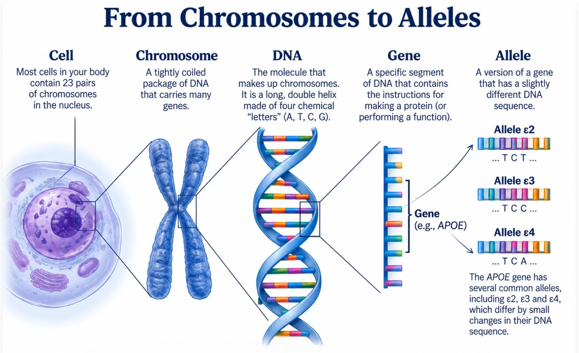
I know this is basic information but I was unaware of how cells and chromosomes worked so I’m sharing it with you. Let’s start at the beginning.
What is a cell?
A cell is the basic living unit of your body. Your body is made of trillions of cells, and although they have different jobs, brain cells send signals, muscle cells contract, and blood cells carry oxygen and they share a basic organization.
Think of a cell as a tiny living factory. It has several parts, each with a specific job.
The Cell and Its Main Parts
Cell membrane — the factory’s outer wall and security gate, controlling what enters and leaves the cell.
Nucleus — the control room, where most of the cell’s DNA is stored.
DNA — the genetic material containing the instructions needed to build and operate the cell.
How DNA Is Organized
Chromosomes — tightly organized packages of DNA that keep the cell’s enormous DNA molecules organized.
Genes — specific sections of DNA located along the chromosomes that contain instructions for making proteins or functional RNA.
Alleles — different versions of a gene. For example, APOE is a gene, while APOE ε2, ε3, and ε4 are different alleles of that gene.
Note: This gets a little confusing,
APOE = the gene
APOE ε2, APOE ε3, APOE ε4 = the alleles/variants of the gene. In some research papers alleles are referred to as APOE2, APOE3, and APOE4.
apoE = the protein; apoE2, apoE3, apoE4 = the three major protein isoforms
From DNA to RNA to Proteins
If you are interested in the actual mechanisms changing DNA messages into proteins, here is the answer.
RNA stands for ribonucleic acid.
RNA is one of the essential molecules in living cells. A simple way to think about the relationship among DNA, RNA, and proteins is:
DNA = the long-term instruction book
RNA = a working copy or messenger that carries and uses some of those instructions. RNA is especially important because it helps turn the information stored in DNA into proteins and also performs other jobs inside cells.
Proteins = the machines and building materials the cell makes from those instructions.
Ribosomes — The Protein-Making Machines (Liu, Kanekiyo, Xu, & Bu, 2013)
Ribosomes — tiny manufacturing machines that read RNA instructions and assemble proteins.
Mitochondria — The Cell’s Power Plants: It is the cell’s power plants, producing much of the energy the cell needs.
Why RNA Matters in Alzheimer’s Research
RNA is especially important in Alzheimer’s research because scientists can study which genes are turned on or off in brain cells and how those patterns change during disease. By examining RNA, researchers can get a picture of what individual cells are actively doing, including changes in the production of proteins involved in inflammation, communication between neurons, energy production, and other processes that may be altered in Alzheimer’s disease. (Murdock & Tsai, 2023) (Mathys & et al, 2019)
APOE
APOE’s Story
The APOE story began not in Alzheimer’s research, but in the study of cholesterol and lipid metabolism. By the early 1980s, scientists had identified three common versions of the APOE gene, ε2, ε3, and ε4, and had begun to understand how their proteins differed. But no one yet knew that one of these genetic variants would become one of the most important clues in Alzheimer’s research. That revelation came in 1993 when APOE ε4 was linked to increased risk of late-onset Alzheimer’s disease.
It wasn’t until the early trials of potential anti-amyloid treatments that it became apparent that ARIA is a risk and a potential side effect of such treatment.
What is APOE?
Like most biological elements ApoE (apolipoprotein E) has a healthy job to do. It acts as a kind of lipid delivery and recycling system. A lipid delivery system is a way the body moves fats (lipids) to and from cells so they can be used, stored, recycled, or removed. This job is especially important in the brain, where lipids are essential for maintaining cell membranes, repairing damaged cells, and supporting the connections between neurons.
In the brain, apoE also interacts with beta-amyloid, influencing how amyloid is transported, aggregated, and cleared. ApoE also helps regulate the brain's immune response. It interacts with microglia, the brain's resident immune cells and influences how they respond to amyloid, damaged cells, and other signals of injury. These are normal biological processes.
The APOE gene has different versions, or alleles; ε2, ε3, and ε4. Each provides instructions for a slightly different form of the apoE protein: apoE2, apoE3, and apoE4.
apoE4
However, the different forms of apoE do not perform these jobs in exactly the same way. In particular, apoE4 can alter microglial responses in ways that, under some conditions, may promote prolonged inflammation and impair the clearance of cellular debris and amyloid that can lead to Alzheimer’s.
Here is what happens:
The APOE gene is a section of DNA located on chromosome 19. (Chromosomes are numbered 1-22, 23 is the sex chromosome either XX or XY)
When a cell needs to produce apoE, it transcribes the APOE gene into messenger RNA (mRNA).
The APOE mRNA carries a copy of the genetic instructions to a ribosome.
The ribosome reads the mRNA and assembles the apoE protein.
If the DNA sequence comes from an APOE ε3 allele, the resulting protein is apoE3. If it comes from an APOE ε4 allele, it produces a protein called apoE4.
One molecule
This is getting into the weeds, but if you are curious about exactly why there is a difference here is an explanation.
The key point is that apoE4 is not a completely different molecule from apoE3. It is almost identical. The difference is caused by one amino-acid substitution in the protein.
The tiny molecular difference
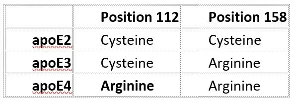
ApoE is a protein made of 299 amino acids. ApoΕ3 and apoE4 differ at just one position:
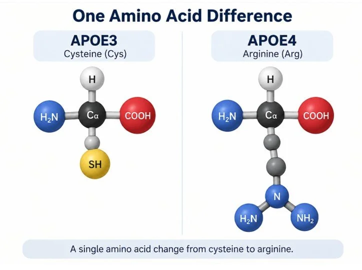
apoE3: amino acid cysteine at position 112
apoE4: amino acid arginine at position 112
That single change from cysteine to arginine changes the protein's chemical properties and affects how parts of the protein interact with each other.
There is also a difference at position 158, but apoE3 and apoE4 both have arginine there:
Why can one amino acid matter so much?
Think of the protein as a folded piece of molecular machinery.
Changing one amino acid can alter the way the protein folds or how different parts of the protein interact. In apoE4, the arginine at position 112 contributes to a different molecular structure and to interactions between regions of the protein that are less prominent in apoE3.
So the important story isn't that apoE4 contains a completely different molecule. It contains one small molecular change that can have consequences for the behavior of the entire protein.
Imagine two delivery trucks built from the same design. Both can carry and deliver packages, but one has a slightly different part in its engine. The truck still works and still delivers its cargo, but the small change affects how efficiently it handles certain loads, how it interacts with the road, or how much wear it experiences over time.
And this is one of the most remarkable aspects of the APOE story: a single change in the sequence of 299 amino acids can alter a protein's behavior enough to influence a person's susceptibility to Alzheimer's disease. (Fouquet, Besson, Gonneaud, La Joie, & Chételat, 2014)
Effects of APOE4
If you are like most of us and you have APOΕ4, you will dwell on the worst case scenario. I hope you don’t. You know when you see an ad on TV for a drug they are required to state all the side effects, many of them quietly ending with “and can cause death”? Well the below are all the worst problems with having an ε4 allele.
Please note: Having APOΕ4 doesn't mean that six biological problems are automatically damaging the brain. Instead, APOΕ4 changes the behavior of the apoE protein in ways that can influence six important biological systems. The effects may be subtle, may differ from person to person, and may change with age and other factors. In some people, these changes may never progress to Alzheimer's disease. In others, they may contribute to the development of amyloid pathology and other changes that eventually increase the risk of Alzheimer's.
How the six changes may—or may not—manifest
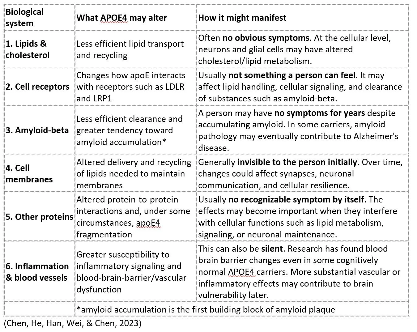
In short: The single amino-acid change in apoE4 can alter the protein's shape and behavior, influencing several systems that help maintain a healthy brain. (Yamazaki, Zhao, Caulfield, Liu, & Bu, 2019)
What APOE combinations mean to you
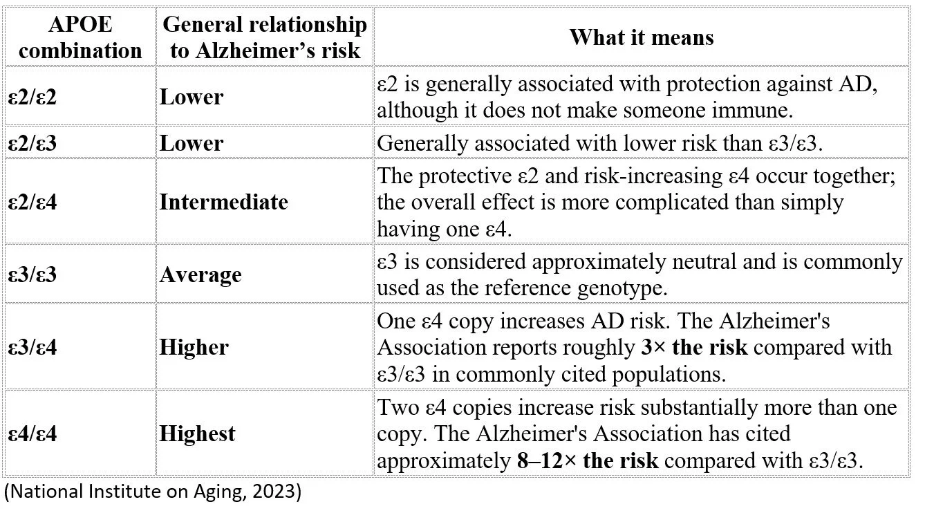
There are six possible APOE combinations, because everyone inherits one APOE allele from each parent: ε2, ε3, or ε4. The combinations are ε2/ε2, ε2/ε3, ε2/ε4, ε3/ε3, ε3/ε4, and ε4/ε4.
APOΕ4 is a risk factor, not a deterministic cause of Alzheimer's disease. It can influence biological processes associated with Alzheimer's, but carrying APOΕ4 does not mean that Alzheimer's will necessarily develop.
ARIA
The Discovery of ARIA
The discovery of ARIA is a good example of how medical progress rarely comes from a single breakthrough. It comes from scientists, physicians, radiologists, drug developers, and clinical-trial participants gradually fitting pieces of a puzzle together.
In the 2000s, researchers were pursuing an ambitious idea: if amyloid-beta was contributing to Alzheimer’s disease, perhaps antibodies could be used to remove it from the brain. One of the first major attempts was bapineuzumab, an experimental monoclonal antibody designed to target amyloid-beta.
The drug ultimately would not become an approved Alzheimer’s treatment, but the trials produced an unexpected discovery that would influence every major anti-amyloid treatment that followed.
Researchers monitoring participants with MRI scans began noticing unusual changes in some patients' brains. Some scans showed areas of swelling or fluid accumulation. Others revealed tiny areas of bleeding. What made the finding particularly intriguing was that many of the people with these MRI abnormalities did not feel ill and showed no obvious symptoms. Something was happening inside the brain that could be seen before it could necessarily be felt.
That observation prompted investigators from multiple institutions to look more closely. Neurologists, radiologists, imaging specialists, clinical researchers, and pharmaceutical scientists compared MRI scans, treatment doses, symptoms, and genetic information from participants in the bapineuzumab trials.
A pattern began to emerge. The abnormalities occurred more often with higher doses of the antibody. Researchers also discovered another important connection: people who carried the APOE ε4 allele, particularly those with two copies, were more likely to experience these abnormalities.
This was an important insight. APOE4 had long been known as a genetic risk factor for Alzheimer's disease, but now it appeared to have another significance, it could also help identify people at greater risk for a complication of amyloid-removing treatment.
Scientists needed a common language for what they were seeing. In 2010, experts working together on these findings introduced the term Amyloid-Related Imaging Abnormalities—ARIA.
Researchers including Reisa Sperling, Stephen Salloway, Nick Fox, David Brooks, Marwan Sabbagh, Lawrence Honig, Anton Porsteinsson and many others continued studying the phenomenon. Their work demonstrated that ARIA was not simply an isolated reaction in a handful of patients. It was a recognizable biological response associated with removing amyloid from the brain. That realization changed the development of anti-amyloid drugs. (Sperling & al, 2012)
The failure of bapineuzumab to become an approved treatment therefore did not make its clinical trials a scientific failure. Those trials left researchers with knowledge that would become extremely valuable when the next generation of antibodies arrived. (Roytman & al, 2022)
By the time lecanemab (Leqembi) and donanemab (Kisunla) were being developed, researchers knew to watch for ARIA.
The story of ARIA illustrates how science advances through accumulated knowledge. An unexpected MRI image in one generation of clinical trials became a clue. Researchers shared and analyzed those clues, recognized a pattern, gave it a name, identified important risk factors, and developed monitoring strategies. The knowledge was then carried forward into the next generation of Alzheimer's treatments.
Today, when someone receiving Leqembi or Kisunla has an MRI to check for ARIA, that scan represents decades of accumulated research and collaboration. And it brings us to the questions that matter most to patients receiving these treatments: What exactly is ARIA? Why does removing amyloid sometimes cause swelling or bleeding? Why does APOE4 increase the risk? And, most importantly, how do physicians detect and manage ARIA while treatment is underway?
Why is it important to know your AOPE?
During the initial test results presented to the FDA for approval of Leqembi and Kisunla ARIA had been identified as a potential side effect. So when these drugs were approved a protocol was put in place to test all prospective patients to determine their risk factor for ARIA. Then proactive testing was put in place to watch for ARIA.
What is ARIA?
ARIA (amyloid-related imaging abnormalities) is a change that can occur in the brain during treatment with amyloid-targeting drugs such as Leqembi (lecanemab) and Kisunla (donanemab). It is called “imaging abnormality” because it is usually detected on an MRI, sometimes before a person has any symptoms. (FDA, 2023)
What causes ARIA?
Some people with Alzheimer’s have amyloid deposited not only in brain tissue but also in the walls of small blood vessels. This is called cerebral amyloid angiopathy (CAA).
When an anti-amyloid antibody begins removing amyloid, the process can affect these amyloid-laden blood vessels. The vessel walls may become temporarily more permeable, allowing fluid or small amounts of blood to escape into surrounding brain tissue.
There are two main forms of ARIA: ARIA-E and ARIA-H
ARIA-E — edema/effusion
The E stands for edema. Fluid leaks from small blood vessels into the surrounding brain tissue, producing swelling or areas of increased fluid on MRI.
It can cause:
headache
confusion
dizziness
nausea
visual disturbances
difficulty walking or coordination problems
seizures in more serious cases
ARIA-E can sometimes produce neurological symptoms that look like a stroke, which is why prompt evaluation is important.
ARIA-H — hemosiderin deposition
The H refers to hemosiderin, a residue left behind when blood has leaked from small vessels.
ARIA-H includes:
microhemorrhages — tiny areas of bleeding
superficial siderosis — blood products deposited along the surface of the brain
less commonly, larger areas of bleeding
Most ARIA is asymptomatic, but serious and, rarely, life-threatening hemorrhages can occur. (FDA, 2023)
The important distinction is between a biological tendency associated with APOΕ4 and something a person actually experiences as a disease or symptom. An APOΕ4 carrier may have some of these changes, several of them, or very little detectable effect for many years. APOΕ4 increases Alzheimer's risk, but it does not mean that a person will necessarily develop Alzheimer's.
Monitoring for ARIA
How Are Patients Who Receive Anti-Amyloid Treatment Monitored?
By the time Leqembi and Kisunla were approved, researchers had learned an important lesson from earlier anti-amyloid trials: ARIA can develop without causing symptoms, so doctors cannot rely on how a patient feels to know whether it is occurring. MRI monitoring therefore became an integral part of treatment, not simply a response to a problem, but a proactive safety system designed to detect ARIA early and give the medical team time to respond.
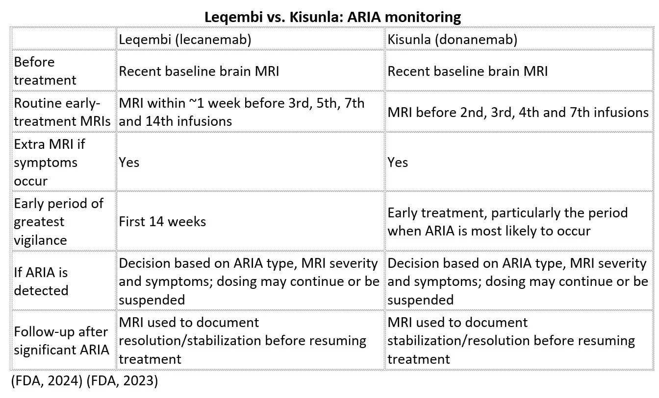
Before treatment begins, the FDA-approved prescribing information for both Leqembi and Kisunla calls for a recent baseline brain MRI. This gives doctors a picture of the brain before treatment and can identify findings such as pre-existing microhemorrhages or superficial siderosis that may increase ARIA risk. Both drugs also call for APOE ε4 testing before treatment to help inform the patient's risk of developing ARIA, particularly because people with two copies of APOE ε4 have a higher risk.
The FDA-approved protocols then build a schedule of MRI examinations into the early part of treatment, when vigilance is especially important. These scans are intended to find ARIA before it necessarily produces symptoms. Patients are also instructed to report symptoms that could indicate ARIA, such as headache, confusion, dizziness, visual changes, or difficulty walking or speaking. When symptoms suggest ARIA, the medical team evaluates the patient and obtains an MRI when indicated.
If an MRI reveals ARIA, the decision is not simply “ARIA equals stop treatment.” Doctors consider several pieces of information together: what type of ARIA is present, how extensive it is on the MRI, and whether the patient has symptoms. Depending on those findings, treatment may continue, be temporarily suspended, or, in more serious circumstances, be discontinued. Follow-up MRI can then show whether the abnormality has stabilized or resolved before treatment is resumed.
This creates a carefully designed early-warning system. The goal is to identify a potential problem as early as possible, assess its seriousness, and give the physician information needed to make the next decision in the patient's best interest.
Leqembi vs. Kisunla: ARIA Monitoring
The two FDA-approved protocols follow the same basic safety strategy, but their routine MRI schedules differ.
Of Note: Kisunla's original dosing gave patients 700 mg at each of the first three infusions before increasing to 1,400 mg. In 2025, the FDA approved a more gradual approach: 350 mg for the first infusion, 700 mg for the second, 1,050 mg for the third, and then 1,400 mg every four weeks. The goal was to remove amyloid more gradually during the early weeks of treatment. In a clinical trial, this change reduced ARIA-E (the form of ARIA associated with brain swelling) while maintaining similar amyloid plaque removal. (Alzheimer's Association, 2025)
Treatment of ARIA
What happens if MRI shows ARIA?
The medical professional who makes the treatment decision is generally the clinician responsible for prescribing and managing the Leqembi or Kisunla treatment; typically a neurologist, geriatrician, or other physician experienced in treating Alzheimer’s with anti-amyloid therapy. The process is usually a team effort, however.
A radiologist/neuroradiologist interprets the MRI. They determine whether the images show ARIA-E (swelling/fluid) and/or ARIA-H (bleeding or blood products), and characterize its severity.
The treating physician reviews the MRI together with the patient's clinical condition.
The doctor considers:
What the MRI shows
Whether the patient has symptoms
How severe the MRI abnormality is
Whether it is ARIA-E, ARIA-H, or both
Previous MRI findings and other risk factors
The treating physician decides whether to continue, temporarily suspend, or permanently discontinue treatment.
The FDA's current Leqembi labeling says that mild, asymptomatic ARIA-E may allow treatment to continue, while moderate or severe ARIA generally calls for suspending treatment. Resumption is guided by clinical judgment after MRI shows resolution and symptoms, if present, have resolved.
Kisunla uses a similar approach: mild, asymptomatic ARIA may allow continued treatment, while more significant or symptomatic ARIA generally results in treatment being suspended until the MRI stabilizes or resolves.
Something to think about
When I first heard the words “ARIA risk,” they sounded frightening because I had no idea what they meant. I didn't understand the connection between a gene I had inherited, the apoE protein it helped produce, and a possible complication of a treatment designed to remove amyloid from my brain. Now I understand that the story is much more complicated—and much more reassuring—than those two words initially suggested. APOE is part of the biology of all of us, APOE ε4 is a risk factor rather than a prediction of what will happen, and ARIA is a known complication that doctors actively look for and know how to monitor and manage. For me, learning the science didn't eliminate the risk; it gave the risk context. And that is what I hope this article does for anyone who hears “ARIA” for the first time: replace fear of an unfamiliar word with enough understanding to have an informed conversation with your doctor about your own treatment.
References
Alzheimer's Association. (2025, April 29). 2025 Alzheimer's disease facts and figures. Retrieved from Alzheimers Association: https://alz-journals.onlinelibrary.wiley.com/doi/10.1002/alz.70235
Alzheimer's Association. (2026). Is Alzheimer's Genetic? Retrieved from Alzheimer's Association: https://www.alz.org/alzheimers-dementia/what_is_alzheimers_%281%29/risk-factors/genetics
Chen, Y., He, Y., Han, J., Wei, W., & Chen, F. (2023, November 12). Blood-brain barrier dysfunction and Alzheimer’s disease: associations, pathogenic mechanisms, and therapeutic potential. Retrieved from Frontiers: https://www.frontiersin.org/journals/aging-neuroscience/articles/10.3389/fnagi.2023.1258640/full
FDA. (2023, July). Leqembi (lecanemab-irmb) injection for intravenous or subcutaneous use initial approval. Retrieved from FDA: https://www.accessdata.fda.gov/drugsatfda_docs/label/2026/761375s001lbl.pdf
FDA. (2024). Kisunla (donanemab-azbt) injections for intravenous use initial approval. Retrieved from FDA: https://www.accessdata.fda.gov/drugsatfda_docs/label/2025/761248s004lbl.pdf
Fouquet, M., Besson, F. L., Gonneaud, J., La Joie, R., & Chételat, G. (2014, August 22). Imaging brain effects of APOE4 in cognitively normal individuals across the lifespan. Retrieved from Springer Nature Link: https://link.springer.com/article/10.1007/s11065-014-9263-8
Liu, C.-C., Kanekiyo, T., Xu, H., & Bu, G. (2013, January 8). Apolipoprotein E and Alzheimer disease: risk, mechanisms and therapy. Retrieved from Nature Reviews Neurology: https://www.nature.com/articles/nrneurol.2012.263
Mathys, H., & et al. (2019, May 1). Single-cell transcriptomic analysis of Alzheimer’s disease. Retrieved from nature: https://www.nature.com/articles/s41586-019-1195-2
Murdock, M. H., & Tsai, L.-H. (2023, July 2). Insights into Alzheimer’s disease from single-cell genomic approaches. Retrieved from Nature Neuroscience: https://www.nature.com/articles/s41593-022-01222-2
National Institute on Aging. (2023, March 1). Alzheimer's Disease Genetics Fact Sheet. Retrieved from National Institute on Aging: https://www.nia.nih.gov/health/alzheimers-causes-and-risk-factors/alzheimers-disease-genetics-fact-sheet
Roytman, M., & al, e. (2022, November 2). Amyloid-Related Imaging Abnormalities: An Update. Retrieved from National Library of Medicine: https://pubmed.ncbi.nlm.nih.gov/36321981/
Sperling, R., & al, e. (2012, March). Amyloid-related imaging abnormalities in patients with Alzheimer's disease treated with bapineuzumab: a retrospective analysis. Retrieved from The Lancet Neurology: https://www.thelancet.com/journals/laneur/article/PIIS1474-4422(12)70015-7/abstract
Yamazaki, Y., Zhao, N., Caulfield, T. R., Liu, C.-C., & Bu, G. (2019, July 31). Apolipoprotein E and Alzheimer disease: pathobiology and targeting strategies. Retrieved from Nature Reviews Neurology: https://www.nature.com/articles/s41582-019-0228-7